Understanding eye health
Corneal cross linking

Written by Dr. Tom Liba
Corneal cross-linking strengthens the cornea to halt keratoconus progression. Who it suits, how it is done and what recovery looks like.

The key points
Briefly
Corneal cross-linking strengthens the cornea to halt keratoconus progression. Who it suits, how it is done and what recovery looks like.
In detail
What is corneal cross-linking and why is it done?
Corneal cross-linking is a treatment meant to stop or slow the progression of keratoconus, a condition in which the cornea thins and bulges forward into a cone shape. It combines riboflavin (vitamin B2) eye drops with ultraviolet A (UVA) light. Together they create new bonds between the collagen fibers of the cornea, making it stiffer and less likely to keep changing shape. The goal is to protect the vision you have and prevent worsening, not to sharpen vision. Vision sometimes improves slightly over time, but this cannot be promised.
Keratoconus treatment: who is it suitable for?
Cross-linking is mainly for people whose keratoconus is progressing, meaning repeated tests show the cornea changing over time. The decision is usually based on comparing corneal topography scans over a period rather than a single test. Teenagers and young adults tend to progress faster, so treatment is sometimes advised relatively early. The cornea also needs to be thick enough, as measured by pachymetry, because a very thin cornea may be damaged by the light. Deep corneal scarring, active infection or very advanced disease may make it less suitable. It is also used in similar conditions where the cornea weakens, for example after laser vision correction. The decision is made individually with an eye doctor.
How is the procedure done?
Treatment is usually done as an outpatient procedure under numbing drops. In the standard method, the thin surface layer of the cornea (the epithelium) is removed so riboflavin can soak in. Riboflavin drops are applied for a while, then the cornea is exposed to UVA light while the drops continue. The whole procedure usually takes under an hour. In another method the epithelium is left in place; recovery may be easier, but its long term effectiveness is still being studied. At the end, a soft bandage contact lens is placed to protect the cornea while it heals.
Recovery after cross-linking
The first few days are usually the hardest. Many people have pain, burning, watering and strong light sensitivity until the surface heals, which typically takes several days. Antibiotic and other drops are used as directed, sometimes with pain relief. The bandage lens is removed at a follow up visit. About a week off work or school is common, and swimming should be avoided until your doctor says it is fine. Vision may be blurry and fluctuate for weeks or even months, and glasses or contact lenses may need refitting. Avoid rubbing your eyes, both during recovery and afterwards.
Side effects and risks
Cross-linking is considered relatively safe, but like any procedure it carries risks. Besides early pain and light sensitivity, mild corneal haze can appear and usually fades over months. Less common problems include slow surface healing, corneal infection and scarring that may affect vision. In some people the disease keeps progressing despite treatment, and repeat treatment or another option may be needed. Reported rates vary between studies and methods, so ask your doctor about the risk in your case.
After treatment: vision, lenses and follow up
Because the treatment mainly stabilizes the cornea, most people still need vision correction afterwards. Glasses or soft lenses may be enough early on, while more advanced cases often use rigid or scleral contact lenses, resumed when the doctor advises. The first check is usually within about a week, followed by further visits over the coming months, and repeat topography confirms the cornea stays stable. If the disease becomes very advanced or significant scarring develops, a corneal transplant may be considered.
When to seek urgent care after treatment
Mild to moderate pain and light sensitivity in the first days are expected. Contact your eye doctor or an eye emergency service right away if pain gets worse after starting to improve, vision drops sharply, there is pus like discharge, redness keeps increasing, or a white spot appears on the cornea. Also call if the bandage lens falls out in the first days. These signs can point to infection, which needs prompt treatment.
More information for you

Eye conditions
Keratoconus: symptoms and treatment
A corneal condition that changes the shape and clarity of vision.
Read more
Eye tests
Corneal topography test
A map of the corneal surface used to detect irregular shape and plan care.
Read more
Eye tests
Pachymetry: corneal thickness test
Pachymetry measures corneal thickness for glaucoma assessment, corneal disease and surgical planning.
Read more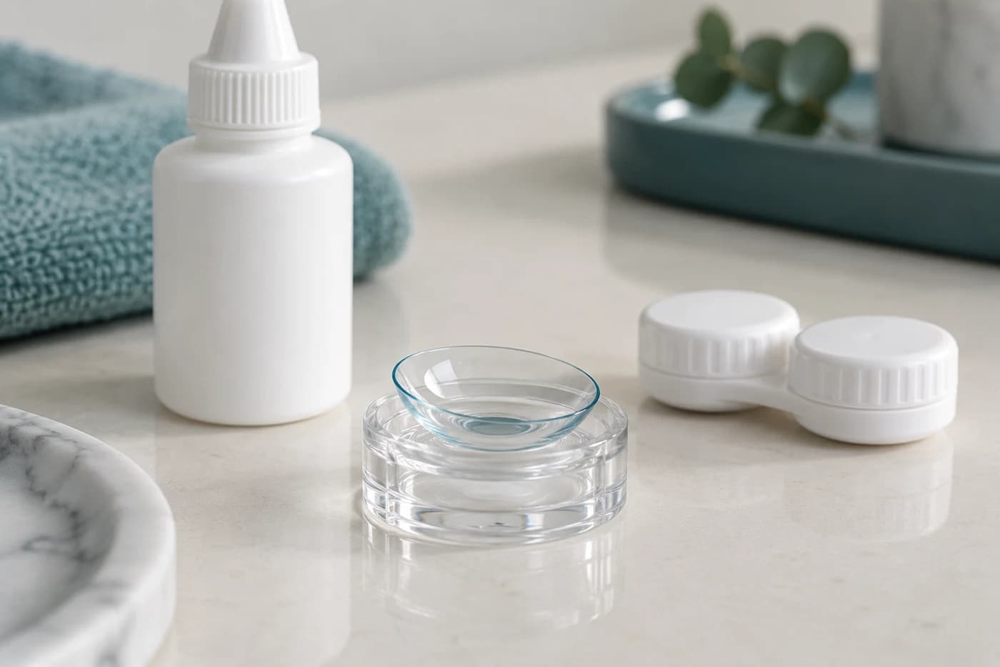
Treatments and surgery
Scleral contact lenses
Large scleral lenses rest on the white of the eye and vault over an irregular cornea.
Read more
Treatments and surgery
Corneal transplant: types and recovery
Transplantation is considered when corneal disease cannot be adequately treated with lenses or medication.
Read moreTom Liba, M.D.
Contact
For questions and further information, you can contact me by email.
tomliba1996@gmail.com