Understanding eye health
Keratoconus: symptoms and treatment

Written by Dr. Tom Liba
A corneal condition that changes the shape and clarity of vision.

The key points
Briefly
Keratoconus is a progressive corneal disease where the cornea thins and bulges into a cone shape, affecting vision quality.
Symptoms include blurred vision, increasing astigmatism, glare, halos, and frequent changes in glasses or contact lens prescriptions.
Treatment options range from glasses and specialized contact lenses to corneal cross linking to halt progression, and corneal transplantation in advanced cases.
In detail
What is Keratoconus?
Keratoconus is a progressive eye condition where the cornea, the clear, dome shaped front surface of the eye, thins and gradually bulges outward into a cone shape. This change in shape distorts the way light enters the eye, leading to blurred and distorted vision. The condition typically begins in adolescence or early adulthood and can affect one or both eyes, often progressing at different rates.
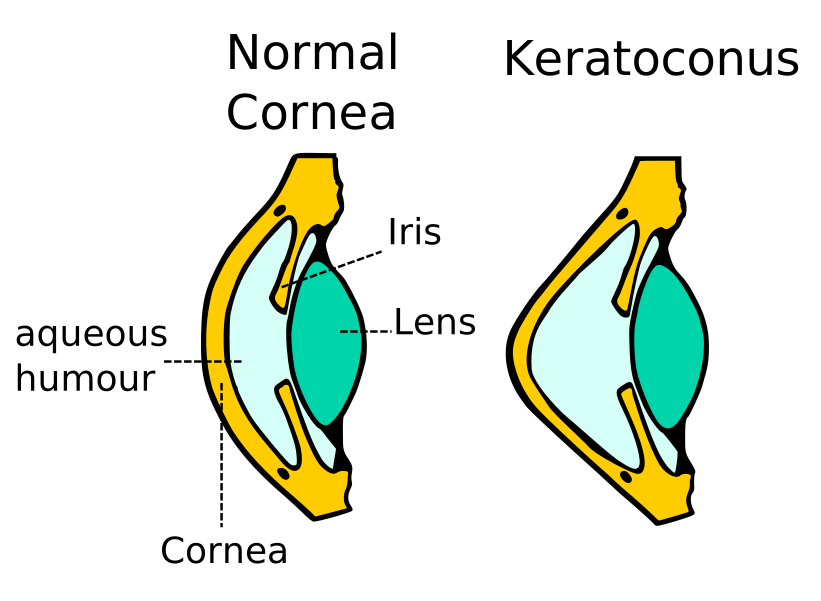
A schematic comparison of a normal cornea and the conical cornea in keratoconus.
Image: Madhero88 · Image source · CC BY SA 3.0 · Used without modification
Symptoms and Risk Factors
Initial symptoms of keratoconus often include mild blurring of vision and increased sensitivity to light. As the disease progresses, individuals may experience significant increases in astigmatism, double vision in one eye, halos and glare around lights, and frequent changes in their glasses or contact lens prescriptions. Risk factors include a genetic predisposition, chronic eye rubbing, and an association with certain allergic conditions.
Diagnosing Keratoconus
Keratoconus is diagnosed through a comprehensive eye examination by an ophthalmologist. This typically involves assessing visual acuity, a slit lamp examination to evaluate the corneal structure, and advanced imaging tests. Corneal topography is a key diagnostic tool that maps the surface curvature of the cornea, identifying changes in its shape and thickness, even in early stages of the disease. These tests allow the doctor to diagnose the condition, assess its severity, and determine its rate of progression.
Vision Correction Options
In the early stages of keratoconus, vision can often be corrected with eyeglasses or soft contact lenses. As the disease progresses and the corneal distortion worsens, specialized contact lenses may be necessary, such as rigid gas permeable (RGP) lenses or scleral lenses. These lenses create a smooth, uniform refractive surface over the irregular cornea, allowing for better vision correction than standard glasses or soft lenses.
Corneal Cross Linking
Corneal cross linking is a procedure aimed at halting the progression of keratoconus by strengthening the corneal tissue. During the treatment, riboflavin (vitamin B2) drops are applied to the cornea, followed by exposure to ultraviolet A (UVA) light. This combination creates new chemical bonds between the collagen fibers in the cornea, making it stronger and more resistant to further bulging. The main goal is to slow or stop progression, rather than to improve vision. Some people experience improved vision over time, but this is not guaranteed, and follow up is needed to check whether the disease remains stable.
Corneal Transplantation
In very advanced cases of keratoconus, when vision is severely impaired and cannot be corrected with contact lenses, or when significant corneal scarring occurs, a corneal transplant may be necessary. In this procedure, a portion or all of the damaged cornea is replaced with healthy corneal tissue from a donor. Various types of corneal transplants exist, and the choice of procedure depends on the severity of the disease and the specific condition of the patient's cornea.
Follow Up and When to Seek Care
Individuals with keratoconus require regular follow up with an ophthalmologist to monitor disease progression and adjust treatment as needed. The frequency of follow up is determined individually. It is important to contact an eye care professional immediately if you experience a sudden and significant decrease in vision, severe eye pain, or unusual redness, as these may indicate complications requiring urgent attention.
More information for you

Tom Liba, M.D.
Contact
For questions and further information, you can contact me by email.
tomliba1996@gmail.com