Understanding eye health
Corneal transplant: types and recovery

Written by Dr. Tom Liba
Transplantation is considered when corneal disease cannot be adequately treated with lenses or medication.

The key points
Briefly
Corneal transplantation is considered when corneal damage cannot be effectively treated with glasses, contact lenses, or medication.
The transplant can involve the entire cornea or specific layers, depending on the disease and anatomy.
Diligent follow up and eye drop use are crucial after transplantation; any change in vision or pain requires urgent assessment.
In detail
What is the Cornea and When is a Transplant Needed?
The cornea is the transparent, outermost layer of the eye, acting as a 'window' through which light enters. It plays a vital role in focusing light rays onto the retina. When the cornea is damaged, it can lose its transparency, leading to a decrease in visual acuity. In situations where vision cannot be adequately improved with glasses, contact lenses, or medication, corneal transplantation may be considered.
Medical Conditions Requiring Corneal Transplantation
Corneal transplantation is necessary for various conditions that affect the cornea's transparency or shape. These include diseases like keratoconus, which causes progressive distortion of the corneal shape, and corneal edema resulting from endothelial cell failure, such as in Fuchs' dystrophy or after complex eye surgeries. Other reasons include corneal scarring from injuries, chemical burns, or infections, as well as rejection of a previous corneal graft or hereditary corneal diseases.
Types of Corneal Transplants
Historically, corneal transplantation typically involved replacing the entire thickness of the cornea. Today, advanced techniques allow for the replacement of only the damaged layers, preserving the healthy ones. This approach reduces risks and shortens recovery time. The type of transplant chosen depends on the location and depth of the corneal damage.
Partial Layer Transplants
Partial layer transplants include DALK, DSEK or DSAEK, and DMEK. DALK (Deep Anterior Lamellar Keratoplasty) replaces the diseased front layers while preserving the patient's healthy endothelium, and may be suitable for advanced keratoconus or anterior scars. DSEK (Descemet Stripping Endothelial Keratoplasty) replaces the damaged inner layers, including the endothelium; DSAEK uses an automated method to prepare similar donor tissue. DMEK replaces only Descemet's membrane and the endothelial cells. Endothelial grafts are used for suitable cases of endothelial failure such as Fuchs dystrophy. Recovery, rejection risk and other complications differ by technique and eye condition, so the surgeon recommends the appropriate approach.
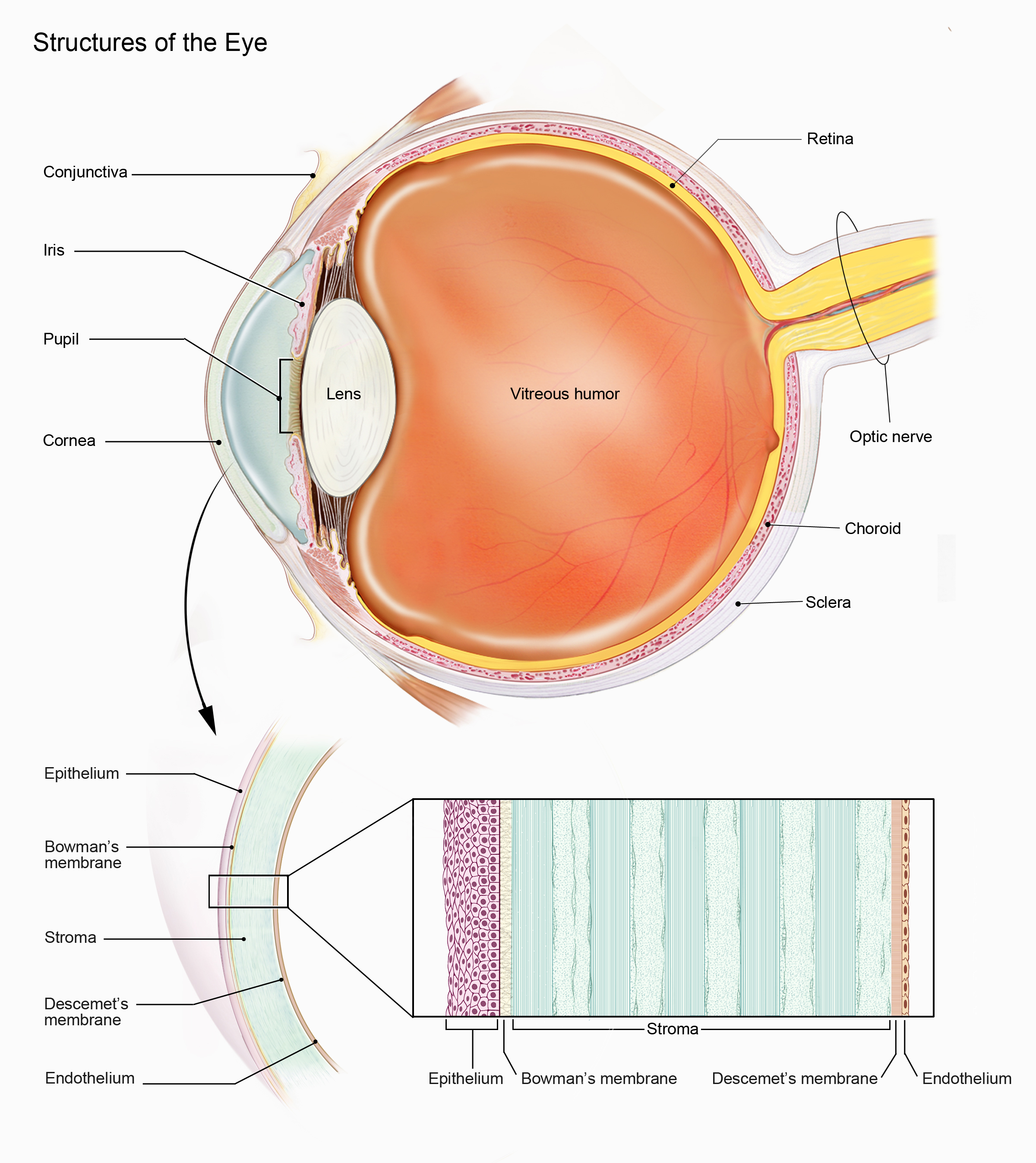
Eye anatomy and corneal layers. Partial transplants replace particular layers according to the affected area of the cornea.
Image: National Eye Institute · Image source · CC BY 2.0 · Used without modification
Preparation for Transplant and Potential Risks
Preparation for a transplant involves a comprehensive evaluation of the eye's condition and overall health. As with any medical procedure, potential risks exist. These include graft rejection, infection, bleeding, changes in intraocular pressure, and significant astigmatism. It is important to discuss all anticipated risks and benefits with your doctor before deciding on the transplant.
Recovery and Post Transplant Care
After transplantation, the recovery period and visual improvement may take some time. It is crucial to consistently use eye drops, often for an extended period, to prevent rejection and infections. Protecting the eye from injury and infection is important, and strenuous physical activity should be avoided in the initial weeks. Regular follow up with an ophthalmologist is essential to monitor the graft's condition and adjust treatment as needed. If an air or gas bubble is used to support an endothelial graft, follow the specific positioning instructions. Do not fly or travel to high altitude until the surgeon confirms it has disappeared. Nitrous oxide must not be used while a bubble remains, so tell any medical, emergency or dental team about the bubble. The surgical team should explain when these restrictions end.
When to Seek Urgent Care
It is important to be aware of warning signs that may indicate a potential complication. You should seek urgent assessment from an ophthalmologist if you experience redness, pain, increased light sensitivity, or a sudden decrease in vision. These signs could indicate graft rejection, infection, or another complication requiring immediate treatment.
Medical sources
More information for you

Eye conditions
Keratoconus: symptoms and treatment
A corneal condition that changes the shape and clarity of vision.
Read more
Eye tests
Corneal endothelial cell count
Endothelial cells help keep the cornea clear; imaging evaluates their density and shape.
Read more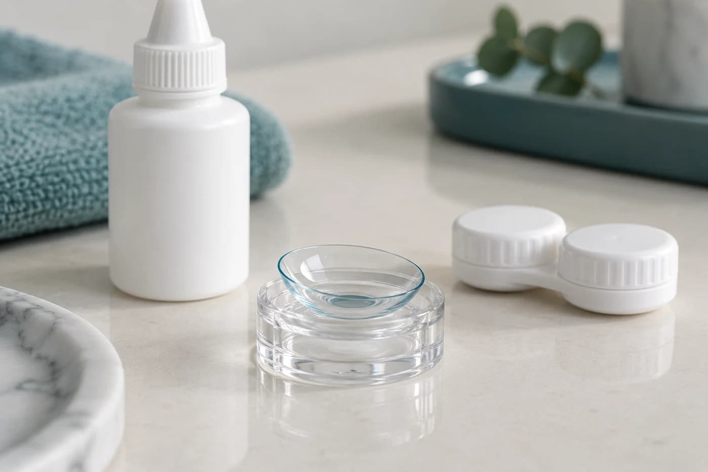
Treatments and surgery
Scleral contact lenses
Large scleral lenses rest on the white of the eye and vault over an irregular cornea.
Read moreTom Liba, M.D.
Contact
For questions and further information, you can contact me by email.
tomliba1996@gmail.com