Understanding eye health
Retinal detachment: symptoms and treatment

Written by Dr. Tom Liba
Retinal detachment separates the retina from supporting tissue.

The key points
Briefly
Retinal detachment is an emergency where the retina separates from its supporting tissue.
Symptoms include flashes of light, new or many floaters, a shadow or curtain in vision, and vision loss.
Treatment may involve laser or cryotherapy for tears, and surgery for detachment.
In detail
What is Retinal Detachment?
The retina is a thin, light sensitive layer lining the back of the eye, functioning much like the film in a camera by converting light into electrical signals sent to the brain. Retinal detachment occurs when this layer separates from the underlying tissue that provides it with nourishment. This is a medical emergency that can cause permanent vision loss if untreated. Seek urgent examination without delay; treatment timing depends on the examination findings and whether the central retina is involved.
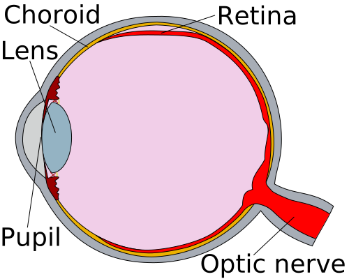
Schematic eye cross section: at the top, the red retina separates from the supporting layer. This simplified diagram is not a patient scan.
Image: Erin Silversmith, Delta G, RexxS · Image source · CC BY SA 3.0 · Used without modification
Causes and Types
Most retinal detachments are caused by a tear or hole in the retina, allowing fluid from the vitreous cavity to pass underneath the retina and separate it. This is known as a rhegmatogenous retinal detachment. Other types include tractional retinal detachment, where scar tissue on the retina pulls it away, and exudative retinal detachment, caused by fluid accumulation under the retina without a tear, often due to inflammation or other diseases.
Risk Factors
Risk factors for retinal detachment include increasing age, high myopia (nearsightedness), posterior vitreous detachment, previous eye surgeries such as cataract surgery, eye trauma, and a family history of retinal tears or detachment. As people age, the vitreous gel filling the eye can shrink and pull away from the retina, a process that can create traction and tears in the delicate retinal tissue.
Symptoms
Symptoms of a retinal tear or detachment may include the sudden appearance of flashes of light (photopsia), a new increase in the number of floaters (small spots or lines drifting in the field of vision), a sensation of a shadow or dark curtain obscuring part of the visual field, or a sudden decrease in visual acuity. These symptoms warrant an urgent examination by an ophthalmologist.
Diagnosis
Diagnosis of retinal detachment involves a comprehensive eye examination, including pupil dilation and examination of the retina using a slit lamp and specialized lenses. In some cases, when the retina cannot be clearly visualized due to hemorrhage or other opacities, an ocular ultrasound may be necessary to assess the retinal status.
Treatment Options
If a retinal tear or hole is detected before detachment occurs, treatment typically involves laser photocoagulation or cryotherapy (freezing treatment). The goal of these treatments is to create a scar around the tear to prevent its progression and subsequent retinal detachment. If retinal detachment has already developed, surgical intervention is usually required. Common surgical procedures include vitrectomy or scleral buckling, sometimes a combination of both, depending on the type and location of the detachment.
Recovery and Follow Up
After laser or cryotherapy, follow up may be needed to ensure proper scar formation and that the retina remains attached. Following surgery, the recovery period varies depending on the type of surgery and the retinal condition. There may be restrictions on physical activity or specific head positioning. It is crucial to follow your doctor's instructions and report any changes in vision or new symptoms. The final visual outcome depends on many factors, including the duration of the detachment and whether the macula (center of vision) was involved.
If surgery leaves an air or gas bubble in the eye, do not fly or travel to high altitude until your surgeon confirms it has fully disappeared. Tell every medical team about the bubble before anesthesia or pain relief. Nitrous oxide, including gas and air used for pain relief, must not be given while the bubble remains because it can dangerously expand the bubble and damage vision.
When to Seek Medical Attention
The sudden onset of flashes of light, a new increase in floaters, a shadow or curtain in your field of vision, or a sudden decrease in vision are warning signs that require immediate examination by an eye doctor. Retinal detachment is an emergency, and you should not wait for a routine appointment. Prompt medical attention can significantly impact the success of treatment and preservation of vision.
Medical sources
More information for you

Eye conditions
Eye floaters and flashes
Eye floaters and flashes are common and usually harmless, but a sudden change can signal a retinal tear. What causes them, and when to seek urgent care.
Read more
Eye conditions
Posterior vitreous detachment (PVD)
Aging vitreous gel may separate from the retina.
Read more
Treatments and surgery
Retinal laser treatment
Retinal laser treats selected tears or abnormal vessels; it differs from refractive laser surgery.
Read more
Treatments and surgery
Vitrectomy: retina surgery
Vitrectomy treats selected retinal detachments, holes, membranes or vitreous hemorrhage.
Read more
Treatments and surgery
Scleral buckle for retinal detachment
A scleral buckle repairs selected retinal detachments based on the tear pattern.
Read moreTom Liba, M.D.
Contact
For questions and further information, you can contact me by email.
tomliba1996@gmail.com