Understanding eye health
Posterior vitreous detachment (PVD)

Written by Dr. Tom Liba
Aging vitreous gel may separate from the retina.

The key points
Briefly
The vitreous is a clear gel filling the eye, which can separate from the retina with age.
This is usually a natural and harmless process, but sometimes it can cause a retinal tear.
New symptoms like flashes of light or floaters require an urgent eye exam to rule out a tear.
In detail
What is Posterior Vitreous Detachment?
The vitreous is a clear, gel like substance that fills the space inside the eye, maintaining its shape and attaching to the retina, the light sensitive tissue at the back of the eye. As people age, the vitreous undergoes natural changes, becoming more liquid and shrinking. This process can cause the vitreous to separate from the retina, a condition known as posterior vitreous detachment (PVD). In most instances, PVD is a benign process and does not cause significant harm to vision.
How PVD Occurs
When the vitreous shrinks and liquefies, it can pull on the retina. Typically, the attachment between the vitreous and retina is weak enough to allow for a smooth separation. However, in some areas, the connection might be stronger, and this pulling can create a tear in the retina. If left untreated, such a tear can lead to retinal detachment, a medical emergency that can result in significant vision loss. Understanding this mechanism is crucial for recognizing potential complications.
Common Symptoms
The most common symptoms of PVD include the sudden appearance of new floaters in the field of vision, which may look like small specks, cobwebs, or rings. Another symptom is flashes of light, typically seen in the peripheral vision, especially in dim lighting conditions. These symptoms are caused by the vitreous pulling on the retina or by shadows cast by the detached vitreous onto the retina. It is important to remember that these symptoms can also indicate a retinal tear, necessitating prompt medical evaluation.
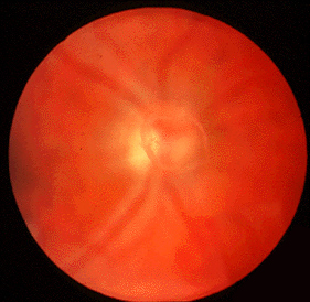
A clinical photograph of vitreous opacities that may be perceived as floaters. An image alone cannot establish whether there is vitreous detachment or a retinal problem.
Image: Jonathan Trobe, MD, University of Michigan Kellogg Eye Center · Image source · CC BY 3.0 · Used without modification
Risk Factors
The primary risk factor for PVD is increasing age, with the condition becoming more common after age 60. Other risk factors include high myopia (nearsightedness), as highly myopic eyes tend to be longer, placing more tension on the vitreous and retina. Eye trauma, previous eye surgeries (such as cataract surgery), and inflammatory eye diseases can also increase the risk of PVD or accelerate its occurrence. These factors contribute to the changes in the vitreous gel.
Diagnosis and Examination
If new symptoms of floaters or flashes of light appear, it is important to seek an eye examination promptly. The ophthalmologist will administer eye drops to dilate the pupils, then perform a thorough examination of the retina using a specialized instrument and lens. This examination allows the doctor to determine if only a PVD is present, or if complications such as a retinal tear or vitreous hemorrhage have developed. Accurate diagnosis is essential for determining the appropriate course of action.
Treatment Options
In most cases, posterior vitreous detachment without an associated retinal tear or other complication does not require specific treatment, and the symptoms tend to improve over time. If a retinal tear is diagnosed, laser treatment or cryotherapy (freezing) may be recommended to seal the tear and prevent the development of retinal detachment. These treatments create a scar around the tear, firmly securing the retina to the eye wall. In rare cases of retinal detachment, surgical intervention may be necessary.
Follow Up and When to Seek Urgent Care
Keep any repeat retinal examinations arranged by your ophthalmologist after a new symptomatic vitreous detachment, even if the first examination found no tear. The timing depends on the findings and your risk factors. Even after an initial examination that finds no complications, it is crucial to remain vigilant for changes in symptoms. Seek immediate medical attention if new or worsening symptoms appear, such as a sudden and significant increase in the number of floaters, the appearance of a 'curtain' or 'shadow' obscuring part of the visual field, a sudden decrease in vision sharpness, or more frequent and intense flashes of light. These symptoms could indicate the development of a retinal tear or detachment, requiring urgent medical intervention.
More information for you

Eye conditions
Eye floaters and flashes
Eye floaters and flashes are common and usually harmless, but a sudden change can signal a retinal tear. What causes them, and when to seek urgent care.
Read more
Eye conditions
Retinal detachment: symptoms and treatment
Retinal detachment separates the retina from supporting tissue.
Read more
Treatments and surgery
Retinal laser treatment
Retinal laser treats selected tears or abnormal vessels; it differs from refractive laser surgery.
Read moreTom Liba, M.D.
Contact
For questions and further information, you can contact me by email.
tomliba1996@gmail.com