Understanding eye health
Watery eye: blocked tear duct

Written by Dr. Tom Liba
Tear drainage pathways carry tears to the nose.

The key points
Briefly
Tear duct obstruction prevents proper tear drainage, leading to excessive tearing, discharge, and increased risk of infections.
Diagnosis involves eye examination, tear drainage tests, and sometimes imaging to identify the location and cause of the blockage.
Treatment varies by age and obstruction site, including massage, dilation, probing, or surgery to create a new drainage pathway.
In detail
The Tear Drainage System and Its Obstruction
Tear glands produce tears that lubricate the eye's surface. These tears drain through small openings at the inner corners of the eyelids, pass through tiny canals into the tear sac, and then descend through a duct into the nose. A blockage anywhere in this drainage system prevents tears from flowing properly. As a result, tears accumulate on the eye's surface and spill over, a condition known as epiphora or excessive tearing.
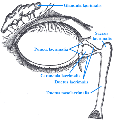
An anatomical illustration of tear production and drainage. Tears pass through small openings in the eyelids to the lacrimal sac and then through the drainage duct toward the nose.
Image: Henry Vandyke Carter, Gray's Anatomy · Image source · Public domain · Used without modification
Symptoms and Complications
The primary symptom of a tear duct obstruction is persistent watering of the eye, often accompanied by mucous or purulent discharge. Stagnant tears in a blocked drainage system can allow bacteria to multiply, increasing the risk of infection in the tear sac or recurrent discharge. An infection of the tear sac (dacryocystitis) can cause painful swelling and redness at the inner corner of the eye.
Risk Factors
Tear duct obstruction can occur at any age. In infants, it is often congenital, resulting from incomplete development of the drainage system or a thin membrane blocking the duct. In adults, risk factors include older age (especially in women), narrowing of the drainage openings, chronic eye inflammation, prolonged use of certain eye drops (e.g., for glaucoma), trauma, infection, tumors, radiation therapy to the face, and certain chemotherapy drugs. Previous surgeries around the eye, eyelids, or nose can also lead to scarring and obstruction.
Diagnosis
Diagnosis begins with a comprehensive eye examination, including inspection of the eye and eyelids. The doctor may perform a tear drainage test by placing a colored drop on the eye's surface and observing how quickly it disappears. Another test involves irrigating the tear ducts with saline solution through the drainage openings to check for fluid passage. In some cases, imaging tests such as X ray, CT, or MRI may be necessary to pinpoint the obstruction's location or identify underlying causes like tumors. An ENT evaluation may also be required to rule out structural abnormalities in the nose.
Treatment Options for Infants and Children
In infants, congenital tear duct obstruction often resolves spontaneously by one year of age as the drainage system matures. A clinician can show parents how to perform tear sac massage with downward pressure at the inner corner of the eye. The appropriate technique matters, and pressure should not be applied to the eyeball. If the obstruction does not resolve, dilation of the drainage openings or probing of the tear ducts may be considered. In rare cases, when these treatments are ineffective, surgery may be necessary.
Treatment Options for Adults
Treatment for adults depends on the location and cause of the obstruction. Narrow drainage openings may be dilated or enlarged surgically with a procedure called punctoplasty. Probing, a temporary tube or balloon dilation may be considered for selected narrowing elsewhere in the drainage pathway. For complete obstruction of the tear sac or nasolacrimal duct, the common treatment is dacryocystorhinostomy (DCR) surgery. This procedure creates a new pathway between the tear sac and the nasal cavity, allowing tears to drain directly into the nose. If a bacterial infection is present, treatment depends on the infection: drops may help surface discharge, but acute infection of the tear sac usually needs systemic antibiotics, taken by mouth or given intravenously in severe cases; however, this treatment does not resolve the obstruction itself.
Follow Up and When to Seek Urgent Care
The follow up and treatment plan are tailored individually for each patient, based on age, type, and severity of the obstruction. It is important to seek urgent medical attention if symptoms such as painful swelling and significant redness at the inner corner of the eye, fever, or rapid worsening of tearing and discharge occur. These symptoms may indicate an acute infection requiring immediate treatment.
More information for you

Treatments and surgery
Tear duct surgery (DCR)
Tear drainage blockage may cause watering or recurrent infection; examination checks other causes of watering..
Read more
Eye conditions
Dry eyes: symptoms, causes and treatment
Common symptoms, risk factors and treatment approaches for dry eye.
Read more
Ophthalmology topics
Oculoplastics: eyelid and tear duct care
Eyelids, tear ducts, brows and the tissues around the eye.
Read moreTom Liba, M.D.
Contact
For questions and further information, you can contact me by email.
tomliba1996@gmail.com