Understanding eye health
Retinal diseases: symptoms and treatment

Written by Dr. Tom Liba
Macular degeneration, diabetic retinopathy, vein occlusion and retinal detachment.

The key points
Briefly
The retina is the light sensitive tissue at the back of the eye, and retinal diseases can affect central or peripheral vision.
Common conditions include macular degeneration, diabetic retinopathy, retinal vein occlusion, and retinal detachment.
A sudden increase in floaters, flashing lights, or a dark curtain in your vision requires immediate eye examination.
In detail
What is the Retina and Its Function?
The retina is a thin layer of nerve tissue lining the back inner surface of the eye. Its primary role is to receive light focused by the lens, convert it into neural signals, and transmit these signals to the brain via the optic nerve. The brain then interprets these signals as the images we see. The macula, located at the center of the retina, is responsible for sharp, central vision, which is crucial for tasks like reading, recognizing faces, and driving. The peripheral retina handles side vision and night vision.
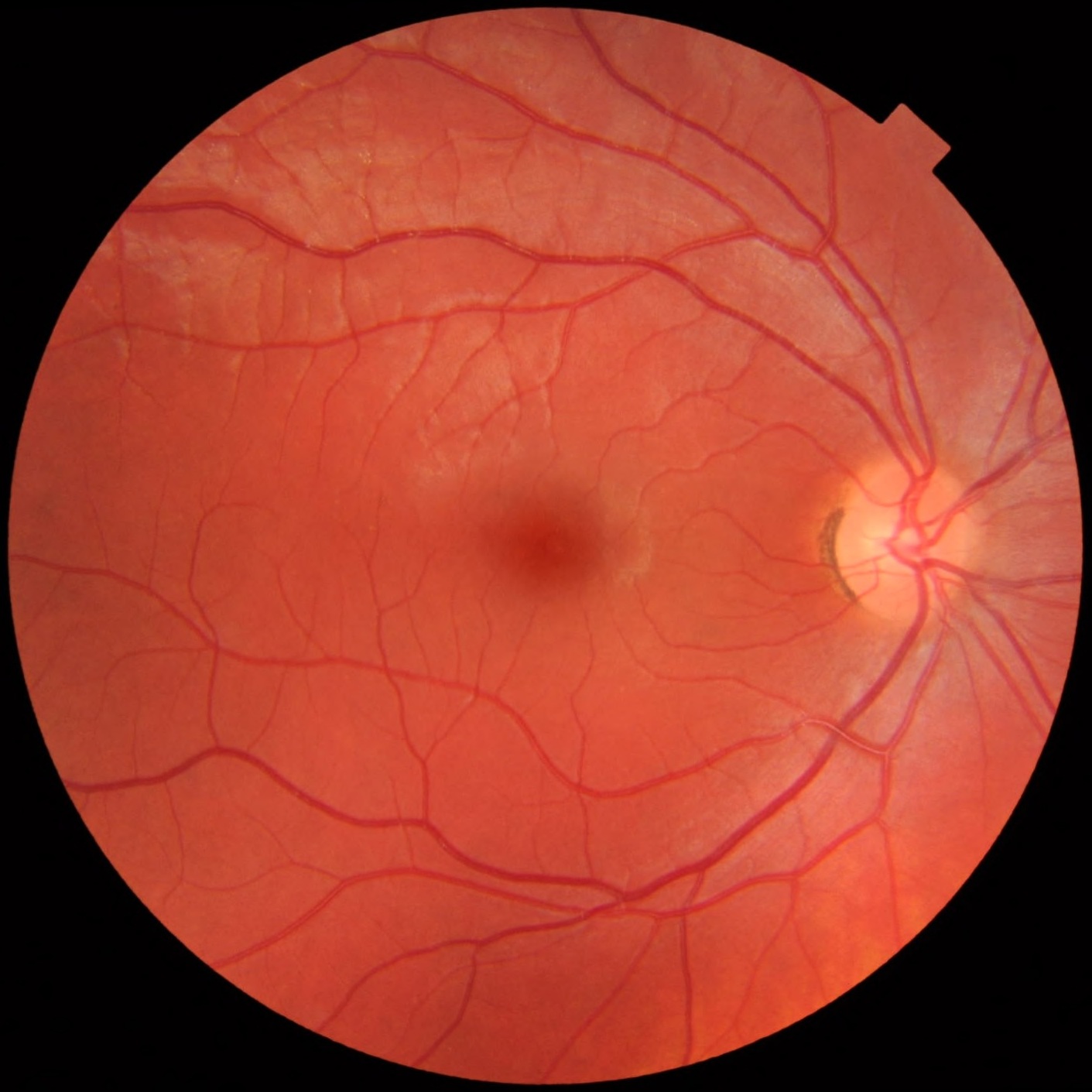
A photograph of a healthy retina. Blood vessels and the optic disc, appearing brighter on the right side of the image, are visible.
Image: Mikael Häggström · Image source · CC0 1.0 · Used without modification
Common Retinal Conditions
Retinal diseases can affect any part of the retina, leading to vision impairment. Age related macular degeneration (AMD) damages the macula, causing central vision loss. Diabetic retinopathy is a complication of diabetes that affects the blood vessels in the retina. Retinal vein occlusions occur when retinal veins become blocked, leading to fluid leakage and hemorrhages. A retinal tear is a break in the retina. Fluid can pass through a tear and separate the retina from the layers beneath it, causing retinal detachment. New symptoms suggesting either condition require urgent examination.
Symptoms and Warning Signs
Symptoms of retinal diseases can include decreased visual acuity, distorted vision (straight lines appearing wavy), blind spots in the field of vision, or difficulty with night vision. Urgent warning signs that require immediate attention from an eye doctor include a sudden increase in numerous floating spots ('floaters'), flashes of light (like a camera flash), or the sensation of a dark curtain descending over your field of vision. These symptoms can indicate a retinal tear or detachment, conditions that require prompt treatment to prevent irreversible vision loss.
Diagnosing Retinal Diseases
Diagnosis of retinal diseases begins with a comprehensive eye examination, including pupil dilation to allow the doctor to view the back of the eye. Additional imaging tests may include Optical Coherence Tomography (OCT), which provides detailed cross sectional images of the retina to identify fluid accumulation or structural changes. Retinal photography (fundus photo) documents the appearance of the retina. In some cases, fluorescein or ICG (indocyanine green) angiography may be performed to evaluate blood flow in retinal vessels and detect blockages. It is important to note that OCTA does not evaluate leakage from blood vessels. Before angiography, inform the medical team about all allergies, especially previous reactions to injected dyes, and about kidney or liver disease, pregnancy or breastfeeding. The team will assess precautions for the particular dye being used. Fasting or forced fluid intake are not universally required, and specific instructions will be provided individually. If breastfeeding, discuss with the medical team regarding the need for temporary interruption. If the retina cannot be seen clearly, for example because of bleeding into the vitreous, an eye ultrasound may help assess its condition.
Treatment Options
Treatment for retinal diseases varies depending on the specific diagnosis. For conditions like wet macular degeneration and diabetic macular edema, intravitreal injections of anti VEGF (vascular endothelial growth factor) medications are a common treatment. These medications reduce fluid leakage and the growth of abnormal blood vessels. Retinal laser treatments may be used to seal leaking blood vessels or treat retinal tears. In cases of retinal detachment, significant vitreous hemorrhage, or macular hole, vitrectomy surgery may be necessary, where the vitreous gel is removed and the retinal issue is addressed. If a gas bubble is placed in the eye during surgery, flying is prohibited as long as the gas remains, and anesthesia staff must be informed before any future surgery due to interaction with nitrous oxide. Specific head positioning should only be maintained if explicitly instructed.
Understanding Risks and Limitations
It is important to understand that despite available treatments, it is not always possible to restore vision to its original state. The goal of treatments is often to stabilize the condition, prevent further deterioration, and preserve existing vision. Every treatment carries potential risks, such as infection, bleeding, or increased intraocular pressure, although these are rare. It is crucial to discuss the benefits and drawbacks of each treatment option and realistic expectations for outcomes with your treating physician.
Follow Up and When to Seek Care
Regular follow up with an ophthalmologist is essential for anyone diagnosed with a retinal disease, even if vision is stable. The frequency of follow up is determined by your doctor based on your specific condition. Do not stop blood thinners or eye drops without explicit direction from your treating physician. If you experience any sudden change in vision, such as a sharp decrease, new distortions, or the sudden appearance of many floaters or flashes of light, you should seek immediate attention from an eye doctor or emergency department. In cases of sudden vision loss, it is crucial to urgently rule out arterial occlusion or arteritis (such as temporal arteritis) which requires immediate treatment. Early detection and treatment can prevent irreversible vision damage.
More information for you

Tom Liba, M.D.
Contact
For questions and further information, you can contact me by email.
tomliba1996@gmail.com