Understanding eye health
Macular hole: surgery and recovery

Written by Dr. Tom Liba
A macular hole affects detailed central vision, often through vitreous traction.

The key points
Briefly
A macular hole is an opening in the macula, the central part of the retina responsible for sharp, detailed vision.
Symptoms include distorted vision, blurriness, or a central blind spot, diagnosed via eye examination and OCT imaging.
Treatment is typically surgical, involving vitrectomy and the injection of a gas bubble into the eye.
In detail
What is a Macular Hole?
A macular hole is a small break or opening in the macula, the central part of the retina. The macula is responsible for sharp, detailed central vision, which is essential for tasks like reading, recognizing faces, and driving. When a hole forms in the macula, it disrupts the eye's ability to see fine details clearly. This condition is distinct from macular degeneration, although both affect the macula.
Causes and Risk Factors
The primary cause of a macular hole is vitreous traction. As we age, the vitreous gel, which fills the center of the eye, naturally shrinks and pulls away from the retina. Sometimes, it remains attached to the macula and exerts a pulling force, eventually creating a hole. Other risk factors include advanced age (over 60), high myopia (nearsightedness) which can thin the retina, eye trauma, retinal detachment, and certain inflammatory conditions.
Symptoms
Symptoms of a macular hole typically develop gradually and affect central vision. They may include blurred vision, distorted vision where straight lines appear wavy or bent (like doorframes or window blinds), and a gray or black spot in the center of the visual field. People often notice difficulty with reading or seeing small details. It is important to note that a macular hole does not usually cause pain.
Diagnosis
Diagnosis of a macular hole involves a comprehensive eye examination. This includes dilating the pupils and examining the retina with a slit lamp. Optical Coherence Tomography (OCT) is a crucial diagnostic tool. OCT provides detailed cross sectional images of the retina, allowing the ophthalmologist to visualize the hole, its size, location, and the extent of vitreous traction, which helps in determining the most appropriate treatment.
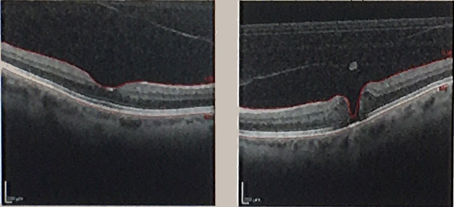
OCT showing a macular hole in the right panel, alongside a comparison section in the left panel.
Image: Betty Wills (Atsme) · Image source · CC BY SA 4.0 · Used without modification
Treatment Options
The main treatment for a macular hole is a surgical procedure called vitrectomy. During the surgery, the vitreous gel is removed to relieve any pulling on the retina. Often, a very thin layer of tissue on the surface of the macula, such as the internal limiting membrane (ILM) or an epiretinal membrane, is gently peeled away to release tension and help the hole close. Finally, a special gas bubble is injected into the eye's cavity. This gas bubble acts as an internal 'tamponade,' gently pressing on the hole to help it close and heal. In rare cases, very small holes may close spontaneously.
Recovery and Expectations
After surgery, specific head positioning instructions may be given for several days or weeks to ensure the gas bubble applies optimal pressure to the hole. This positioning is not necessarily face down and will be tailored individually. Vision will be very blurry as long as the gas bubble is present in the eye, gradually improving as the gas absorbs and is replaced by the eye's natural fluid. This process can take several weeks to months. The degree of visual improvement depends on factors such as the hole's size, its duration before surgery, and individual healing. It is important to understand that perfect vision, as it was before the hole developed, may not be fully restored.
Post Surgery Precautions
If a gas bubble has been injected into the eye, it is absolutely forbidden to fly in an airplane or travel to high altitudes while the gas is present. Changes in atmospheric pressure can cause the gas bubble to expand, leading to a dangerous increase in intraocular pressure. Any medical team, especially an anesthesia team, must be informed about the presence of gas in the eye before any other medical procedure, as nitrous oxide can cause dangerous expansion of the gas bubble. It is crucial to follow all post operative medical instructions, including the use of eye drops and follow up examinations.
Follow up and When to Seek Care
After surgery, regular follow up appointments will be scheduled to ensure the hole has closed properly and vision is improving. It is important to attend all scheduled follow up visits. You should seek immediate ophthalmological attention if you experience severe eye pain, significant redness, a sudden and sharp decrease in vision, or the appearance of flashes of light or a 'curtain' in your field of vision, as these could indicate potential complications.
More information for you


Tom Liba, M.D.
Contact
For questions and further information, you can contact me by email.
tomliba1996@gmail.com